 EN
EN
 FR
FR
 ES
ES
 AR
AR

Biomechanical Optimization for Osteoporotic Bone and Minimally Invasive Fixation
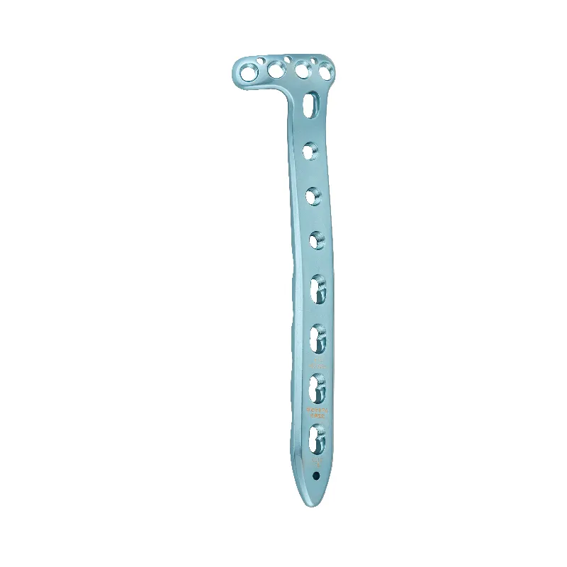
When designing a distal femoral plate for minimally invasive surgery, engineers face some pretty tough tradeoffs. The main issues are dealing with how fragile osteoporotic bones really are, plus working around the fact that surgeons have very limited space to operate. Getting this right means finding that sweet spot between making sure the implant stays stable mechanically and letting the body heal naturally. We want to encourage those early callus formations without tearing up too much soft tissue or creating problems from stress shielding. It's a delicate dance between engineering requirements and what biology can actually handle during recovery.
Balancing torsional rigidity and axial micromotion: Tapered cross-sections and variable screw density
When axial micromotion stays under about half a millimeter, it actually helps stimulate callus formation around bones. Some recent studies from last year showed patients with osteoporotic fractures healed about three weeks faster when this level of controlled movement was maintained during treatment. Another important design feature involves tapered cross sections which help reduce those pesky stress concentrations right at the screw holes where failures often occur in weakened bone structures. Looking at screw placement patterns, we find that having more screws concentrated near the fracture area while reducing them further down the bone can boost torsional strength by roughly thirty percent without affecting blood flow through the cortex. These approaches work together to maintain structural stability while still allowing for natural bone healing processes, something that makes perfect sense when considering modern minimally invasive surgical techniques focused on smaller incisions and quicker patient recoveries across orthopedic practices today.
Preventing stiffness mismatch: Material selection (e.g., titanium alloys) and porous surface integration
Titanium alloys have become the go to choice for those low profile distal femoral plates mainly because they match cortical bone better than stainless steel does. Their Young's modulus sits around 40 percent closer to what we see in real bone tissue, which means there's less stress shielding going on and the load gets transferred more naturally across the implant site. When combined with those porous surfaces that actually boost the strength at the bone implant interface by roughly a quarter thanks to better osseointegration, these materials really tackle the problem of stiffness mismatch head on. What all this adds up to is much better long term stability when dealing with osteoporotic bones. This matters a lot during percutaneous procedures where getting good initial grip and maintaining that hold over time makes all the difference between success and failure in surgical outcomes.
Percutaneous Screw Placement Accuracy: Locking Mechanism and Angle Optimization
Fixed-angle vs. polyaxial locking: Trade-offs in targeting flexibility and construct stability
The type of locking mechanism selected makes a big difference for what happens during surgery as well as how patients recover afterward. Fixed angle designs basically stop any movement between screws and plates, which gives them really good resistance against twisting forces. This matters a lot when dealing with bones that aren't very dense because even tiny movements at the fracture site can slow down healing time. But these fixed systems require extremely accurate planning before surgery starts and often need lots of X-ray guidance during placement. On the other hand, polyaxial systems allow about 15 degrees of adjustment while inserting screws, making it easier to work around different body shapes and getting better results through small skin incisions. They aren't quite as stiff as fixed angle ones though, showing roughly 12 to 18 percent less strength when compressed repeatedly. Still, studies indicate that polyaxial options cut down on the need for repeat surgeries caused by misplaced screws by approximately 27 percent. There's no one size fits all solution here. Surgeons have to consider factors like the specific break pattern, how strong the surrounding bone is, and their own level of expertise with each technique.
±15° convergence envelopes and screw trajectory guides: Reducing fluoroscopy dependence
The design includes convergence envelopes set at around ±15 degrees from the plate axis along with physical guides for trajectories. Together these allow surgeons to place bicortical screws reliably through small stab incisions instead of needing big cuts in soft tissue or constantly checking with fluoroscopy. What makes this work so well is how the system actually limits where drills can go, which helps avoid breaking through cortex and increases chances of getting it right on the first try. When looking at screw arrangements validated by finite element analysis, we see load distribution across six key spots in the bone. Clustering screws near both ends of fractures gives extra stability there, whereas spacing them out in the middle part maintains blood supply and keeps stress from building up too much in one spot. According to clinical simulations, over 92% of attempts succeed on the first pass when using this method. Plus, each screw takes about 40% less time under fluoroscopy compared to traditional freehand approaches. Studies have also shown that this particular arrangement of six screws provides maximum stability during mechanical tests, making it a solid choice for many surgical applications.
Surgical Workflow Integration: Jigs, Indirect Reduction, and MIS-Specific Instrumentation
Getting surgical workflows to work together smoothly isn't just something extra it's actually really important for making sure MIS distal femoral plating works safely and effectively. Specialized jigs help place screws accurately through small incisions and can cut down on fluoroscopy usage by around 40% when compared to older methods according to research published in the Journal of Orthopaedic Trauma last year. The instruments need to fit different body shapes and sizes while still working with regular MIS tool kits that surgeons already have. For fixing fractures, doctors often use indirect methods instead of directly manipulating bones. This approach relies on things like ligament tension and special adjustable devices to line up broken bones properly. The big plus here is that this technique doesn't strip away the outer layer of bone tissue which helps maintain blood flow critical for healing especially in patients with weak bones from osteoporosis.
Leading systems now incorporate three key ergonomic innovations:
- Single-use trajectory guides with built-in ±15° angulation limits to prevent errant screw trajectories
- Magnetic coupling between jigs and plates for stable, hands-free positioning in confined surgical corridors
- Low-friction screwdriver interfaces that minimize soft-tissue snagging and torque-related complications
Collectively, these features reduce average procedure time by 25 minutes and improve procedural reproducibility across training levels (Clinical Biomechanics, 2023). The outcome is more predictable fixation, shorter operative windows, lower radiation exposure, and reduced iatrogenic soft-tissue injury–translating directly into faster patient recovery and improved resource utilization.
FAQ Section
What are the main challenges in designing distal femoral plates for osteoporotic bones?
Designers must balance between mechanical stability of the implant and allowing natural healing while dealing with the fragility of osteoporotic bones and the limited space available for surgery.
What role does titanium play in treating osteoporotic fractures?
Titanium alloys are used in distal femoral plates because they better match the mechanical properties of bone, reducing stress shielding and encouraging natural bone healing.
How does screw placement affect healing in osteoporotic bone surgery?
Optimally distributing screws near fracture areas enhances torsional strength without harming blood supply, fostering better healing in fractures.
What is the difference between fixed-angle and polyaxial locking systems?
Fixed-angle systems provide sturdy resistance against twisting but require precise placement, while polyaxial systems offer flexibility in screw placement at the cost of some stability.
Why is surgical workflow integration important in minimally invasive surgeries?
Smooth integration helps ensure the success and safety of surgeries by reducing operation time, radiation exposure, and tissue injury, thereby aiding quicker recovery.
Table of Contents
- Biomechanical Optimization for Osteoporotic Bone and Minimally Invasive Fixation
- Percutaneous Screw Placement Accuracy: Locking Mechanism and Angle Optimization
- Surgical Workflow Integration: Jigs, Indirect Reduction, and MIS-Specific Instrumentation
-
FAQ Section
- What are the main challenges in designing distal femoral plates for osteoporotic bones?
- What role does titanium play in treating osteoporotic fractures?
- How does screw placement affect healing in osteoporotic bone surgery?
- What is the difference between fixed-angle and polyaxial locking systems?
- Why is surgical workflow integration important in minimally invasive surgeries?