 EN
EN
 FR
FR
 ES
ES
 AR
AR

Anatomical and Growth-Specific Constraints of the Distal Medial Tibia
Metaphyseal Flare, Epiphyseal Proximity, and Their Impact on Plate Contouring and Screw Placement
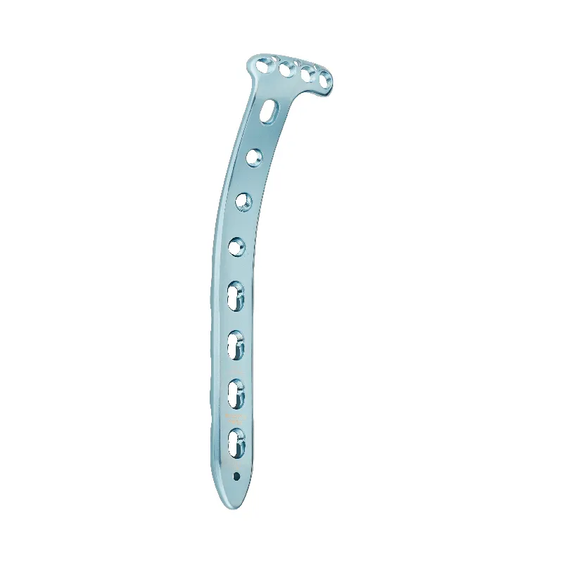
The distal medial tibia features an inverted trumpet-shaped medullary cavity—transitioning from thick cortical bone proximally to thin cortex with abundant cancellous bone distally. This metaphyseal flare presents three key challenges for pediatric plate design:
- Contouring plates to match the ~45° slope at the flare junction
- Avoiding screw placement within 5 mm of the physis to prevent growth arrest
- Accommodating a 40% reduction in cortical thickness at the metaphysis-diaphysis junction versus mid-diaphysis
Successful fixation requires plates with 15–25° variable-angle screw options—enabling safe bypass of the physis while preserving subchondral support in Salter-Harris III/IV fractures.
Age-Related Changes in Tibial Geometry: From Neonatal Bowing to Skeletal Maturity
Tibial morphology evolves across four developmental phases, each demanding tailored fixation strategies:
| Age Range | Key Structural Change | Plate Design Implication |
|---|---|---|
| 0–3 years | Anterior bowing (15° avg) | Pre-contoured 10° dorsal bend |
| 4–8 years | Medial malleolar ossification | Distal hook extension for epiphyseal capture |
| 9–13 years | Physeal width reduction (3.2 – 1.1 mm/year) | Transition to hybrid screw–epiphyseal wire fixation |
| 14+ years | Medial torsion increase (5° – 23°) | Anti-rotational screw trajectory guides |
Concurrently, the distal tibia’s cross-section shifts from triangular to circular during adolescence—necessitating plates with ~30% reduced thickness at skeletal maturity to avoid soft-tissue impingement.
Growth-Preserving Engineering Principles in Pediatric Orthopedic Plate Design
Low-Profile Construction (<1.2 mm) and Edge Optimization to Minimize Soft-Tissue Irritation
The thinner layers of soft tissue in children create real problems with implants sticking out and causing irritation or breaking down the skin. We've found that plates thinner than 1.2 mm work much better, particularly when they have those nice rounded and tapered edges. These are especially helpful around areas where bones stick out prominently, think about places like the lower part of the shin bone. Get these plates shaped just right and they lie flat against the flared part of the bone without losing their strength or affecting how the child grows naturally. This balance between protection and allowing normal development is what makes these designs so valuable in pediatric cases.
Material Selection: Titanium Alloys for Biocompatibility, MRI Compatibility, and Corrosion Resistance
Ti-6Al-4V titanium remains the go to material for pediatric plates because it strikes just the right balance between being safe inside the body, works well during MRIs, and stands up to corrosion over time. The fact that it's not magnetic means doctors can get clear images without interference from metal parts showing up on scans. Plus, since it resists corrosion so well, there's much less chance of harmful metal ions getting released into growing bones, which is especially important around areas where bones are still developing. All these characteristics together make sure the implants stay safe and work properly for years, even as children grow and change.
Biomechanically Informed Plate Configuration for Small-Bone Fracture Fixation
Medial vs. Anterolateral Placement: Stability, Physeal Safety, and Surgical Access Trade-offs
Plate positioning for distal medial tibia fractures involves balancing three interdependent priorities:
- Stability: Medial placement offers superior compressive strength via direct cortical contact—but increases physeal risk in the narrow metaphyseal flare zone.
- Physeal Safety: Anterolateral placement increases distance from the physis by 15–20%, reducing growth disturbance risk—yet compromises torsional rigidity due to lower bone density in that region.
- Surgical Access: Anterolateral approaches reduce soft-tissue complications by ~30% and avoid neurovascular structures; medial access allows more precise anatomical adaptation but carries higher technical demand.
Triaxial optimization—prioritizing physeal preservation without sacrificing fixation integrity—enables 95% retention of natural growth potential in small-bone fracture fixation.
Clinical Evidence and Future Directions in Pediatric Orthopedic Plate Design
Real-World Outcomes: Multi-Center Study of Distal Medial Tibia Plates in Salter-Harris III Fractures
In a recent 2023 study across multiple centers looking at 112 cases of Salter-Harris III fractures, researchers found that about 96 out of every 100 patients showed proper bone healing on X-rays within just eight weeks when treated with specially shaped, low profile plates. Only around 3 percent experienced any issues with growth disturbance during this period. These findings really back up what many orthopedic surgeons have been seeing clinically regarding how important it is to position screws carefully around the growth plates and optimize the edges of implants to prevent both tissue irritation and damage to growing bones. When compared with older techniques, the new approach where screws are placed with greater precision cuts down the chance of bones shifting again after surgery by roughly 40 percent. Looking ahead, there's exciting work happening in biodegradable implants that eventually dissolve in the body, which means no need for another operation to remove metal parts later on. Some companies are also developing compression systems that actually adjust as children grow, tackling some long standing problems we've had with treating fractures in smaller bones.
FAQs on Pediatric Orthopedic Plate Design
What are the main challenges in designing plates for pediatric tibia fractures?
Pediatric tibia fractures pose unique challenges such as contouring plates to fit the metaphyseal flare, avoiding growth arrest, and accommodating reduced cortical thickness at the metaphysis-diaphysis junction.
Why is Ti-6Al-4V titanium preferred for pediatric plates?
Ti-6Al-4V titanium is favored for its biocompatibility, MRI compatibility, and corrosion resistance, ensuring safety and effectiveness in growing children.
How do age-related changes in tibial geometry affect plate design?
Plate design must adapt to age-related changes such as anterior bowing, medial malleolar ossification, and medial torsion increase, requiring tailored fixation strategies for different age groups.
Table of Contents
- Anatomical and Growth-Specific Constraints of the Distal Medial Tibia
- Growth-Preserving Engineering Principles in Pediatric Orthopedic Plate Design
- Biomechanically Informed Plate Configuration for Small-Bone Fracture Fixation
- Clinical Evidence and Future Directions in Pediatric Orthopedic Plate Design
- FAQs on Pediatric Orthopedic Plate Design