 EN
EN
 FR
FR
 ES
ES
 AR
AR

Additive Manufacturing: Driving Customization and Biomechanical Optimization
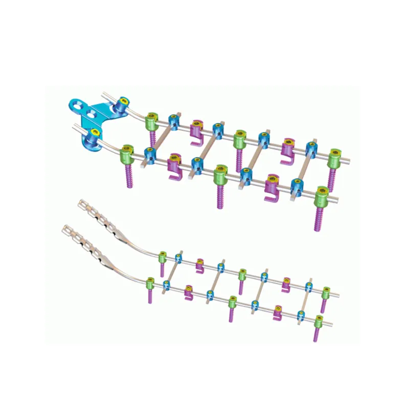
3D-Printed Porous Titanium Cages for Enhanced Osseointegration and Anatomic Fit
With additive manufacturing, surgeons can now create spinal cages tailored specifically for each patient's unique anatomy, featuring controlled porosity gradients that actually address individual anatomical differences while encouraging new bone growth. The titanium lattice structures used in these cages closely resemble the natural cancellous bone density we see in healthy spines, with pore sizes ranging around 300 to 700 micrometers. This design increases the available surface area for cells to attach by roughly 40 to 60 percent when compared to standard solid implants. What makes these cages really stand out is their interconnected pore network, which helps blood vessels grow through them and allows osteoblasts to migrate more effectively. Clinical tests have shown this leads to faster fusion rates, improving results by about 25 to 30 percent. When the cage geometry matches the shape of the vertebral endplate precisely, it reduces problems like stress shielding and subsidence risks. This becomes especially important in complicated spinal deformities where conventional cages often don't fit properly. Surgeons are increasingly seeing better long term outcomes thanks to this technology that lets them design implants based on what biology actually needs rather than what was possible with older manufacturing methods.
Comparative Overview of DMLS, SLS, and SLA in Spinal Implant Fabrication
| Process | Materials | Resolution | Key Advantages | Primary Limitations |
|---|---|---|---|---|
| DMLS (Direct Metal Laser Sintering) | Titanium alloys | 20–40 µm | High-density load-bearing parts; enables complex lattice geometries impossible via machining | Post-processing requirements; residual stress management |
| SLS (Selective Laser Sintering) | PEEK, TPU polymers | 50–100 µm | Radiolucent, flexible interbody devices for motion preservation | Limited control over pore architecture and mechanical consistency |
| SLA (Stereolithography) | Biocompatible resins | 10–25 µm | Exceptional surface fidelity for surgical guides and anatomical models | Not suitable for permanent load-bearing implants due to polymer degradation and fatigue limitations |
Each process requires validation specific to its layer-by-layer fabrication risks—including residual stresses, powder contamination, and thermal distortion—especially for permanent implants subject to long-term biomechanical loading.
Smart Implants and Digital Integration for Postoperative Intelligence
Embedded Strain/Load Sensors Enabling Real-Time Recovery Monitoring
Spinal implants equipped with built-in strain and load sensors represent a major change in how we approach recovery after surgery. These devices constantly track the mechanical stresses acting on the hardware, giving doctors immediate information about how well bones are fusing together and whether the implant remains stable. Doctors can spot problems early on, like strange movements or too much pressure on certain areas, which might lead to implant failure or poor bone healing if left unchecked. This lets them intervene sooner with specific treatments before things get worse. Rehabilitation plans aren't just generic anymore either. Patients receive personalized activity recommendations that change over time based on actual tissue responses instead of sticking rigidly to predetermined schedules. The wireless feature means doctors can monitor patients remotely through secure apps, cutting down on redundant doctor visits while keeping patients more engaged in their own recovery. Some preliminary studies indicate that following recovery paths guided by sensor data could cut down on the need for repeat surgeries considerably.
Balancing Clinical Value with Regulatory and Data Management Challenges
Getting smart implants into widespread use really comes down to solving these big regulatory issues plus building proper data infrastructure. The problem is that regulatory bodies want proof not just about how safe the physical implant is, but also about whether the digital parts work reliably, stay secure from cyber threats, and actually deliver medical benefits. This creates a whole different approval process compared to regular medical devices. At the same time, all those sensors constantly sending data need storage that meets HIPAA standards, full encryption from start to finish, and they have to connect smoothly with existing electronic health records. Hospitals are struggling too with figuring out what signals matter most so doctors don't get buried under meaningless notifications. Making progress here will take everyone working together across industries - manufacturers making the implants, doctors who will use them, IT folks handling the data side, and government regulators. They need to build systems where different technologies can talk to each other while keeping patient information private, which isn't easy when trying to speed up innovation without putting anyone at risk.
Next-Generation Biomaterials and Bioactive Surface Engineering
Titanium, PEEK, and Functionally Graded Materials in Motion-Preserving Devices
Today's motion preserving spinal implants are moving toward materials like titanium alloys, PEEK plastic, and these newer things called functionally graded materials (FGMs). These materials help balance how well the implant works mechanically with how it integrates into the body. Titanium is strong but light, and we know it bonds well with bone tissue over time. PEEK has another advantage though - it doesn't show up on X-rays as much, which makes monitoring easier for doctors. Plus, its stiffness matches real bone pretty well, cutting down on stress problems by about 15 to 30 percent compared to older metals like stainless steel or cobalt chrome. Functionally graded materials are really interesting because they change properties across different layers. Think of something that's hard titanium inside but has a softer, porous surface coated with hydroxyapatite. This mimics the way our own spine tissues naturally vary in density. All these improvements support better stabilization systems that keep normal movement between vertebrae while reducing wear on nearby segments. Looking at actual results from clinics, patients who got FGM based cervical discs reported being satisfied around 92% of the time after two years, which speaks volumes about both how well these implants work and how long they last before needing replacement.
Bioactive Coatings to Accelerate Fusion
Surface engineering techniques, especially those involving hydroxyapatite (HA) coatings and controlled release of bone morphogenetic protein-2 (BMP-2), really boost spinal fusion outcomes. These methods work by attracting osteoblasts and encouraging mineral deposits where they're needed most. Studies show HA coatings can increase bone implant contact by around 40 to 60 percent within just three months through a process called biomimetic mineralization. When BMP-2 delivery is properly optimized, fusion happens about 30 to 50 percent faster compared to implants without any special coatings. But there's a catch worth mentioning here. If too much BMP-2 gets released, we see problems like unwanted bone growth in more than 20 percent of patients, which often means going back for another surgery. That's why new dual layer coatings are making waves in the field right now. They first release antimicrobials like silver or gentamicin to fight infections, then gradually introduce growth factors that help regenerate tissue exactly where it should be. For lumbar fusions specifically, these improved surfaces have cut down on healing time from an average of nine months down to just six months. Patients get back to normal activities sooner, which makes all the difference in their quality of life after surgery.
Surgical Enablement: Robotics, Navigation, and Intraoperative Imaging Synergy
The combination of robotics, navigation tech, and intraoperative imaging has become a game changer for spine surgery accuracy. During operations, doctors can get live, detailed images of anatomy thanks to CT or MRI scans right in the operating room. These navigation systems take that image data and turn it into moving 3D guides for instruments, making sure screws go in the right spot over 90% of the time. Then robotic arms carry out these plans with incredible precision at the millimeter level, which means smaller cuts and less damage to surrounding tissues. Surgeons still have complete control throughout the whole procedure, feeling what's happening through special feedback systems in their tools. All together, these technologies cut down bleeding during surgery by around 30%, get patients out of the hospital faster, and reduce serious complications from nerve damage, especially when dealing with complicated spinal issues. What we're seeing isn't just another small step forward, but really marks a major shift in how minimally invasive spine surgery works today. Precision, safety, and consistency aren't just nice to have anymore they're built into each part of the surgical process now.
FAQ
What is additive manufacturing in spinal surgery?
Additive manufacturing in spinal surgery refers to the process of creating customized spinal implants using 3D printing technology and controlled porosity gradients to match individual patient anatomy and encourage new bone growth.
How do smart implants work?
Smart implants are equipped with embedded sensors that track mechanical stresses on the hardware, offering real-time data on bone fusion and implant stability, allowing for early detection of potential issues.
Why are next-generation biomaterials important?
Next-generation biomaterials like titanium alloys and PEEK are crucial for spinal implants as they offer better mechanical performance, integration into the body, and reduce stress shielding, enhancing patient outcomes.
What are bioactive coatings in spinal implants?
Bioactive coatings, such as hydroxyapatite and BMP-2, accelerate spinal fusion by attracting osteoblasts and initiating mineralization, significantly reducing healing time.
How do robotics and imaging improve spine surgery?
Robotics and imaging technologies provide detailed live anatomical images and guide instruments with precision, reducing surgical complications and improving accuracy in spine surgeries.