 EN
EN
 FR
FR
 ES
ES
 AR
AR

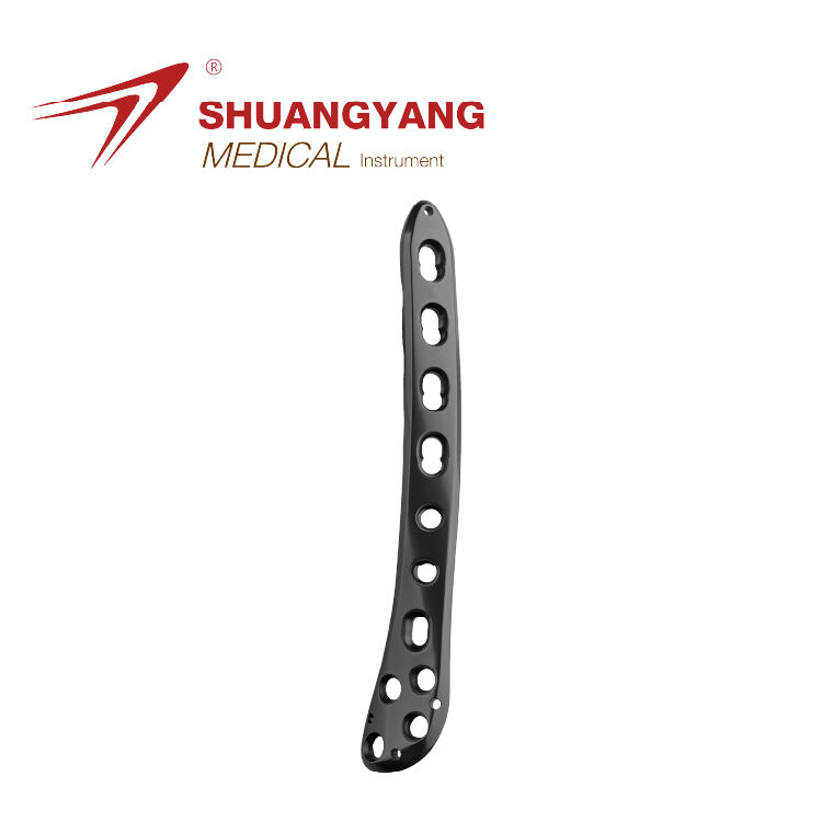
Understanding Dimensional Accuracy in Patient-Matched Tibia Plates
Defining Dimensional Accuracy in Patient-Specific Osteosynthesis Plates
When we talk about dimensional accuracy for those custom made tibia plates, we're basically looking at how well the implant matches up with the actual shape of someone's lower leg bone. The goal is to keep any differences under half a millimeter because anything bigger than that can cause problems with how it fits inside the body and supports movement properly. If manufacturers go over this tiny margin, patients face higher chances of issues down the road. For instance, screws might come loose in some cases, something studies show happens around 28% more often when there's poor fit according to research published in the Journal of Orthopaedic Trauma last year. Plus, healing takes longer too since pressure gets distributed unevenly across the bone. Generic implants just don't cut it here. Custom plates actually take into account all sorts of personal differences in bone structure including curves and thicknesses of different layers. Most importantly, these details get spotted through CT scans and 3D models about 9 out of 10 times when dealing with complicated tibial fractures, as noted in International Orthopaedics earlier this year.
The Role of CT Imaging and 3D Reconstruction in Capturing Anatomical Precision
CT scans that have a slice thickness of about 0.625 mm along with pixel resolution around 0.35 by 0.35 mm create very detailed digital images of the distal tibia's shape. These scans pick up on important anatomical features like the lateral malleolar groove and where the anterior tibial tendon runs through - things that get overlooked in nearly two thirds of surgical planning cases when doctors just look at regular X-rays according to research published in BMC Musculoskeletal Disorders last year. Using this kind of data, medical teams can now print out real life sized models of patients' bones. Surgeons actually try fitting plates onto these models before going into surgery which cuts down on having to make changes during operations by almost half. Plus it helps them line up screws exactly where they need to go relative to how weight naturally distributes across the bone structure.
Clinical Consequences of Dimensional Inaccuracy in Custom Plates
Implant-Bone Interface Mismatch and Its Impact on Fracture Reduction
Tiny deviations from perfect alignment can really mess up the fit needed for proper fracture fixation. According to a recent study published in the Journal of Bone & Joint Surgery last year, when surgical plates make contact with about 92% of the bone surface area, doctors see around a 41% drop in cases where bones fail to heal properly compared to plates that only touch 78% of the surface. When implants aren't aligned right, they tend to move slightly against the bone, which creates extra stress points especially around where the screws go in. Research from the Biomechanics Institute back in 2022 showed this stress increases between 18 and 22 percent in these areas. Some surgeons try to fix this problem by tightening their screws too much, but this actually speeds up bone loss and can ruin all the hard work done during the initial surgery to get everything lined up correctly.
Screw Placement Errors Due to Plate Misfit and Alignment Issues
Surgical guides made specifically for patients using CT scans cut down on drilling mistakes by about two thirds when compared to doing it manually according to research published in the Annals of Orthopedic Surgery back in 2021. Even minor mistakes matter though. An angle off by just 2.1 degrees might move where screws go by nearly 4 millimeters at the bone surface, which is enough to make things unstable in almost 3 out of every 10 situations. Looking at cadaver tests reveals something interesting too. Plates that follow ISO 12891 size requirements hit the mark with screw placement around 98 percent of the time, while those that don't meet these standards only manage about 73 percent accuracy. This shows clearly why getting measurements right matters so much for keeping everything stable after surgery.
How 3D Printing and CAD/CAM Enable Anatomically Accurate Plate Design
Modern orthopedic solutions leverage 3D printing and Computer-Aided Design/Manufacturing (CAD/CAM) to deliver unmatched dimensional accuracy in patient-matched distal tibia plates, bridging the gap between anatomical complexity and surgical execution.
CAD/CAM Integration in Preoperative Planning for Distal Tibia Fractures
Computer aided design and manufacturing systems take CT scans of anatomy and turn them into detailed three dimensional models, which lets surgeons create custom plates accurate to within about half a millimeter. The whole process allows doctors to tweak things like how curved the plate needs to be and where the screw holes should go before surgery even starts. This cuts down on those tough decisions during actual operations by almost 60 percent when compared with old fashioned methods. When looking at broken bones, these systems analyze the fracture patterns too, helping place implants in spots that spread out pressure evenly across all the broken pieces. Makes sense really, nobody wants their hardware failing because it wasn't positioned right.
Validating the Accuracy of 3D-Printed Models in Surgical Implant Design
Coordinate measuring machines (CMM) are used to verify 3D-printed prototypes against digital designs, with clinical studies confirming 98.4% surface congruence in titanium alloy plates. Surgeons report 72% fewer alignment corrections during implantation when using validated patient-specific systems, significantly shortening operative time and enhancing bone-to-implant contact—key factors in promoting fracture healing.
Evidence from Clinical Applications and Case Studies
Case study: Improved surgical precision using CT-based, 3D-printed guides
In a recent study across multiple centers involving 67 patients suffering from complicated distal tibia fractures, researchers found something quite remarkable. When using custom plates created through CT scans and 3D printed guides, doctors needed to make far fewer adjustments during surgery—about half as many alignment corrections compared to traditional methods. The surgical teams noted another benefit too: operations took around 28% less time overall. What's really impressive is that the fit between implants and bones remained extremely precise, with errors staying below 0.8 mm throughout the trials. That kind of accuracy makes all the difference in preventing complications later on. These findings highlight just how valuable these new planning technologies can be when it comes to turning detailed imaging into successful surgeries in actual practice settings.
Measured outcomes in screw placement accuracy with patient-matched plates
Recent findings from the Journal of Orthopedic Trauma (2024) illustrate the clinical benefits of dimensional precision:
| Metric | Patient-Matched Plates | Standard Plates |
|---|---|---|
| Optimal screw placement | 94% | 68% |
| Post-op malalignment | 3% | 19% |
| Revision surgeries | 1.2% | 8.7% |
These improvements stem from CAD/CAM workflows that align screw trajectories within ±0.5° of preoperative plans, minimizing risks of cortical perforation seen in 23% of conventional procedures while maintaining mechanical performance comparable to established 3.5 mm plating systems.
FAQ Section
What is dimensional accuracy in patient-specific osteosynthesis plates?
Dimensional accuracy refers to how closely the custom-made tibia plates match the actual shape and structure of the patient's lower leg bone, ideally within a 0.5 mm difference.
Why is CT imaging crucial in creating patient-matched tibia plates?
CT imaging provides detailed digital images of the bone structure, capturing anatomical precision that helps in designing plates that fit accurately and mitigate surgical risks.
How does 3D printing contribute to the development of custom tibia plates?
3D printing, along with CAD/CAM technology, allows for the creation of anatomically accurate patient-specific plates, improving fit and reducing operative adjustments.
What are the benefits of using patient-specific tibia plates over standard plates?
Patient-specific plates offer improved screw placement accuracy, reduced post-operative malalignment, and a decreased need for revision surgeries compared to standard plates.